Ralstonia solanacearum race 3 biovar 2
(Printer friendly version)
Figures only
| Author: | Patrice G. Champoiseau of University of Florida |
| Reviewers: | Caitilyn Allen of University of Wisconsin; Jeffrey B. Jones, Carrie Harmon and Timur M. Momol of University of Florida |
| Publication date: | September 12, 2008 |
| Project title: | Ralstonia solanacearum race 3 biovar 2: Detection, exclusion, and analysis of a select agent pathogen |
| Supported by: | The United States Department of Agriculture - National Research Initiative Program (2007-2010) |
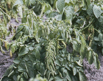 |
Photo 1a. Symptom of brown rot of potato caused by R. solanacearum showing wilting of youngest leaves of plant (Photo courtesy of D.P. Weingartner – IFAS, University of Florida, Hastings) |
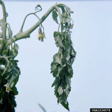 |
Photo 1b. Symptom of bacterial wilt of tomato caused by R. solanacearum showing wilting of leaves at the end of plant branch (Photo courtesy of Clemson University - USDA Cooperative Extension Slide Series, Bugwood.org) |
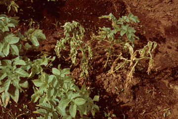 |
Photo 2a. Symptom of brown rot of potato caused by R. solanacearum showing wilting and stunting of plant (Photo courtesy of David Thurston, Cornell University) |
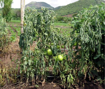 |
Photo 2b. Symptom of bacterial wilt of tomato caused by R. solanacearum showing wilting of foliage and stunting of plant (Photo courtesy of C. Allen, University of Wisconsin) |
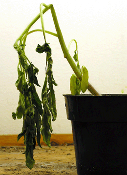 |
Photo 3. Symptom of bacterial wilt of tomato caused by R. solanacearum showing collapse of young stem after artificial inoculation of the plant (Photo courtesy of P. Champoiseau, University of Florida) |
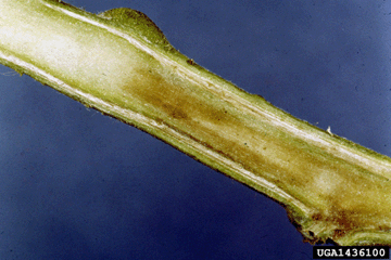 |
Photo 4. Brown discoloration of stem tissues caused by R. solanacearum (Photo courtesy of Clemson University - USDA Cooperative Extension Slide Series, Bugwood.org) |
 |
Photo 5. Grey-brown discoloration of vascular tissues and bacterial ooze in
potato tuber infected
by R. solanacearum (Photo courtesy of K. Tsuchiya) |
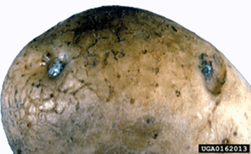 |
Photo 6. Bacterial ooze exuding from eye of potato tuber infected by
R. solanacearum (Photo courtesy of Central Science Laboratory, Harpenden Archive, British Crown, Bugwood.org) |
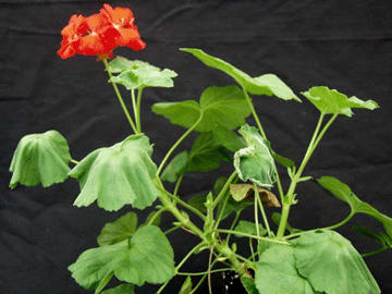 |
Photo 7. Initial symptoms of Southern wilt of geranium caused by R. solanacearum showing wilting and upward curling of leaves (Photo courtesy of D. Norman, Mid-Florida Research and Education Center, IFAS, University of Florida) |
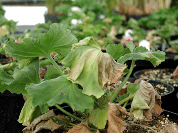 |
Photo 8. Symptoms of Southern wilt of geranium caused by R. solanacearum race 3 biovar 2 showing drying and brown necrosis on leaves (Photo courtesy of the Wisconsin Department of Agriculture, Trade and Consumer Protection) |
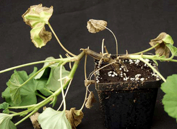 |
Photo 9. Late symptoms of Southern wilt of geranium caused by R. solanacearum race 3 biovar 2 showing blackening and collapse of stem (Photo courtesy of D. Norman, Mid-Florida Research and Education Center, IFAS, University of Florida) |
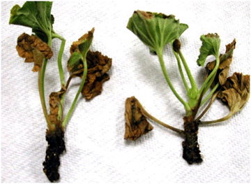 |
Photo 10. Late symptoms of Southern wilt of geranium caused by R. solanacearum showing root blackening (Photo courtesy of Margery Daughtrey, Cornell University) |
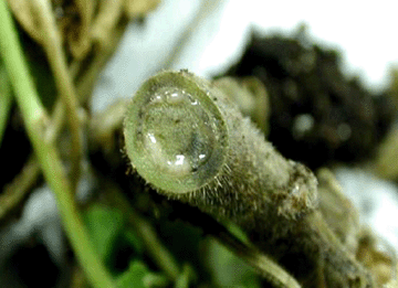 |
Photo 11. Bacterial ooze from freshly cut section of a geranium stem infected by R. solanacearum (Photo courtesy of M. Daughtrey, Cornell University) |
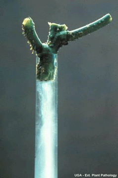 |
Photo 12. Bacterial streaming in clear water from stem cross-section of plant infected by R. solanacearum (Photo courtesy of University of Georgia, Plant Pathology Extension) |
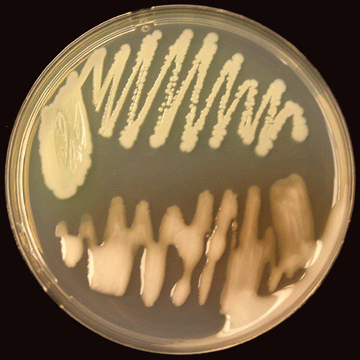 |
Photo 13. Virulent (bottom) and non-virulent (top) colonies of R. solanacearum on CPG agar growth medium (Photo courtesy of P. Champoiseau, University of Florida) |
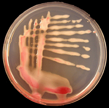 |
Photo 14. Virulent colonies of R. solanacearum on TZC agar medium (Photo courtsey of P. Champoiseau, University of Florida) |
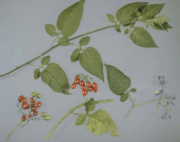 |
Photo 15. Bittersweet or woody nightshade (Solanum dulcamara). Key features for identification (Photo courtesy of J. Elphinstone, Central Science Laboratory, York, UK, Crown Copyright) |
 |
Photo 16. Bacterial streaming in clear water from stem cross-section of plant infected by R. solanacearum (Photo courtesy of University of Georgia, Plant Pathology Extension) |
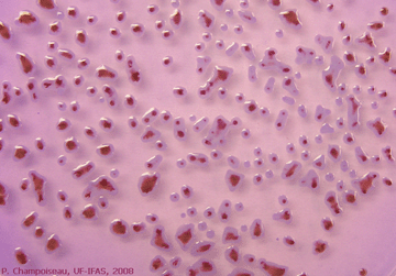 |
Photo 17. Appearance of virulent colonies of R. solanacearum on modified SMSA medium (Photo courtsey of P. Champoiseau, University of Florida) |
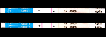 |
Photo 18. Result of a quick serological test showing negative (-) and positive (+) detection of R. solanacearum (Photo courtsey of P. Champoiseau, University of Florida) |
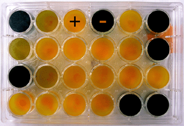 |
Photo 19. Result of a biovar test showing positive (+) and negative (-) utilization of sorbitol by several strains of R. solanacearum (Photo courtsey of P. Champoiseau, University of Florida) |
This R. solanacearum/Bacterial wilt - R3b2 page has been optimized for print. To view this page in its original form, please visit: http://plantpath.ifas.ufl.edu/rsol/Trainingmodules/RalstoniaR3b2_PrinterFigures.html
©2008 R. solanacearum race 3 biovar 2 USDA-NRI project. All rights reserved.